From the desk of a PhD researcher and author of technical papers
For centuries, the human brain has remained biology’s ultimate black box. Its staggering complexity, locked away inside the skull, has made it profoundly difficult to study. Researchers have long relied on imperfect proxies: animal models that don’t fully capture human neurobiology and post-mortem human tissue that offers only a static snapshot of a once-living system. This has left our understanding of complex neurological disorders like autism, schizophrenia, and Alzheimer’s disease frustratingly incomplete.
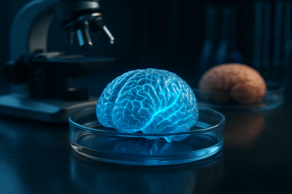
Now, a groundbreaking achievement from a team at Johns Hopkins University is prying that box open. By growing and assembling the first functional, multi-region “mini-brains” in a lab, scientists are creating an unprecedented window into the earliest stages of human brain development and disease. This advance doesn’t just promise to accelerate research; it heralds a new era in drug discovery, personalized medicine, and our fundamental understanding of the mind itself.
A New Blueprint for the Brain
The breakthrough (https://pubmed.ncbi.nlm.nih.gov/40625223/), introduces what the researchers have termed a multi-region brain organoid, or MRBO. To appreciate the significance of this development, one must first understand the limitations of its predecessors.
Beyond Single-Region Models
For years, scientists have been able to grow organoids—three-dimensional clusters of tissue derived from stem cells that self-organize to mimic an organ. While revolutionary, most brain organoids were limited to replicating a single, isolated brain region. As lead author Dr. Annie Kathuria, an assistant professor in JHU’s Department of Biomedical Engineering, explains, “Most brain organoids that you see in papers are one brain region, like the cortex or the hindbrain or midbrain”.
This posed a fundamental problem. Many of the most challenging neurological and psychiatric conditions are not diseases of a single part, but of the connections between parts. “Diseases such as schizophrenia, autism, and Alzheimer’s affect the whole brain, not just one part of the brain,” Kathuria notes. To truly model these disorders, researchers needed a system where different brain regions could grow, connect, and communicate.
An Engineering Feat: Assembling the MRBO
The Johns Hopkins team solved this challenge with an ingenious engineering approach. Instead of relying on cells to self-organize from a single mass, they took a modular approach.
- Grow the Parts: Using human induced pluripotent stem cells (iPSCs)—which can be created from a person’s skin or blood cells and coaxed into becoming any cell type—the team first grew separate cultures of cells representing the three main parts of the brain: the cerebrum, the midbrain, and the hindbrain. Critically, they also grew complex endothelial organoids, which are precursors to a vascular system, complete with a diverse mix of cell types that form rudimentary blood vessels.
- Fuse Them Together: The researchers then took these individual components and fused them using what they describe as a “biological superglue”—a set of sticky extracellular matrix proteins that encourage the tissues to adhere and integrate.
As the separate parts meshed, something remarkable happened: they began to form connections and communicate, functioning as a single, integrated network. This shift from passively growing organoids to actively building them, or creating “assembloids,” represents a new philosophy in bioengineering, one that offers far greater control and complexity.
A Functional Glimpse of Early Life
The true power of the MRBO lies not just in its structure, but in its function. The assembled mini-brain shows clear signs of life, exhibiting biological activity and a startling fidelity to an early-stage human brain.
The Spark of a Network
Once fused, the MRBOs “started producing electrical activity and responding as a network”. This is the crucial evidence that the different regions are not merely co-existing but are functionally interconnected. Scientists can observe this activity using several techniques. One method involves culturing the organoids on microelectrode arrays (MEAs), plates embedded with tiny electrodes that can detect the electrical chatter of neurons. Another, known as calcium imaging, uses fluorescent dyes that cause neurons to “glow” when they fire, allowing researchers to visually track the flow of information across the network.
This coordinated activity is not random. Research on other organoids shows that their electrical patterns mature over time, following a developmental trajectory that closely mimics that of a preterm human infant’s brain waves (EEG). This suggests the MRBO is following a genetically encoded program, making it an invaluable model for studying what happens when that program goes awry.
A Blueprint of the Fetal Brain
The MRBO provides a remarkably accurate snapshot of early human neurodevelopment. Analysis shows that its structure and cellular makeup resemble that of a 40-day-old human fetus.
Table 1: The Multi-Region Brain Organoid (MRBO) at a Glance
| Feature | Specification | Significance |
| Model Name | Multi-Region Brain Organoid (MRBO) | First model to integrate multiple distinct brain regions with a complex vascular system. |
| Core Components | Cerebral, Mid-Hindbrain, and Endothelial Organoids | Allows for the study of communication and connectivity between brain regions, crucial for whole-brain disorders. |
| Assembly Method | Fusion of pre-differentiated components via “biological superglue” (ECM proteins) | Represents an “engineering” or “assembloid” approach, offering more control than pure self-organization. |
| Biological Analogy | 40-day-old human fetal brain (Carnegie Stages 12-16) | Provides an unprecedented window into the earliest stages of human neurodevelopment. |
| Cellular Fidelity | Contains ~80% of cell types found in the developing fetal brain | High-fidelity model that includes diverse neuronal and glial cells, validated by single-nucleus RNA sequencing. |
| Neuronal Scale | 6-7 million neurons per organoid | Significantly smaller than an adult brain (~86 billion neurons) but complex enough to form functional networks. |
| Key Functionality | Spontaneous, coordinated electrical activity across the network | Demonstrates that the fused regions are not just co-located but are functionally interconnected. |
| Vascularization | Includes rudimentary blood vessels and an early blood-brain barrier (BBB) | Critical for modeling nutrient supply, waste removal, and drug delivery across the BBB. |
The inclusion of a vascular system and a nascent blood-brain barrier is particularly vital. It means the MRBO isn’t just a collection of nerve cells; it’s a more holistic system that models how the brain receives nutrients and protects itself, which is essential for testing how drugs might behave in a real human body.
A Revolution in Medicine on the Horizon
With a functional, human-relevant model in hand, researchers can now tackle some of the biggest challenges in medicine, from understanding the roots of brain disease to designing better drugs.
A New Paradigm for Drug Discovery
The development of drugs for neurological disorders is notoriously difficult and expensive. An estimated 96% of neuropsychiatric drugs that look promising in animal models fail once they reach human clinical trials. This catastrophic failure rate is largely because animal models are often poor predictors of human response. As JHU professor and organoid pioneer Dr. Thomas Hartung has famously stated, “we are not 150-pound rats”.
MRBOs offer a powerful solution. By providing a human-cell-based testing ground that more closely resembles the brain, they can help scientists identify which drugs are likely to fail much earlier in the process, saving billions of dollars and years of wasted effort. This could dramatically improve the odds of getting effective treatments to the patients who need them.
Personalized Neurology
Perhaps the most exciting application is the potential for personalized medicine. Because MRBOs can be grown from any individual’s cells, scientists can create a “brain-in-a-dish” that is genetically identical to a patient. This opens the door to a future where doctors can test various treatments on a patient’s personal organoid to see which works best for their unique biology. As Dr. Kathuria envisions, this will allow medicine to “watch disorders develop in real time, see if treatments work, and even tailor therapies to individual patients”. This moves neurology away from a one-size-fits-all approach and toward truly precision-based care.
The Future and Its Ethical Frontiers
The implications of this technology extend even beyond medicine. Some researchers are exploring the concept of “Organoid Intelligence” (OI), aiming to harness the brain’s incredible computational power to create a new form of biological computer.
Of course, as these models become more complex, they raise profound ethical questions. While researchers are clear that current organoids are not conscious, the rapid pace of advancement demands a proactive ethical framework. Recognizing this, the Johns Hopkins team has embedded a consortium of scientists, bioethicists, and public members to help navigate this complex terrain, ensuring that the science progresses responsibly.
The development of the multi-region brain organoid is more than just a scientific curiosity. It is a foundational technology that provides a powerful new tool to unravel the mysteries of the brain, a new platform to cure its most devastating diseases, and a new lens through which we must consider the ethical boundaries of science itself. The assembled mind in the lab dish is just the beginning.


